Pioneering trial reveals alternatives when mammograms fall short in breast cancer detection
Dense breast tissue can mask cancer in conventional tests. A study shows the most effective complementary imaging techniques for detecting malignant lesions in screening programs
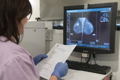

Mammography has become one of the most important tools for early cancer detection, but it may fall short for a certain group of women: those with dense breast tissue — a characteristic that can obscure tumors on mammographic images
There is ongoing debate in the scientific community about how best to address a critical gap in breast cancer screening — cases where conventional mammography falls short. Women with dense breast tissue often escape detection, as their fibroglandular tissue can obscure tumors on mammograms. But questions remain: Which supplemental test is most effective? And which subgroup of women with dense breasts should receive it?
A pioneering clinical trial has now added new data to the discussion. Published Wednesday in The Lancet, the study compared the performance of three complementary imaging tests and found that two contrast-enhanced techniques were more effective than ultrasound when used alongside mammography. However, the research did not evaluate key clinical outcomes, such as whether these methods reduce cancer mortality or the long-term risks of overdiagnosis — so the debate continues.
Breast density is unrelated to breast size. Instead, it refers to the proportion of fibrous and glandular tissue compared to fat. This characteristic is invisible to the naked eye and must be assessed through a mammogram, where fat appears black and dense tissue appears white. The challenge is that tumors also appear white, and in dense breasts, the abundance of fibrous tissue can mask cancerous lesions.
Experts note that women with high breast density face two distinct risks: they are up to four times more likely to develop breast cancer, and their tumors are more likely to go undetected until later stages.
While mammography has a general sensitivity of 85% to 90%, that figure can drop to 60% in women with dense breasts, says Marina Álvarez, a breast imaging specialist with the Spanish Society of Medical Radiology (SERAM). That decline in detection is a key limitation in screening programs, as roughly 10% of women undergoing early detection tests fall into the dense breast category.
How to address these technical limitations remains an open question within the scientific community. There is no clear consensus on how to manage women with dense breast tissue, and the benefits of complementary imaging techniques are still under debate.
In practice, Álvarez explains, there are two scenarios: “In diagnosis, in a breast unit and with women with a history of cancer or at higher risk, we usually follow up the studies with ultrasound. But when it comes to screening, there are no recommendations for complementary tests. There is a lot of debate.”
There’s also a lot at stake. “Because of this lack of sensitivity, cancer can go undetected,” says Álvarez, who is also director of the Radiodiagnosis and Breast Cancer Unit at the Reina Sofía University Hospital in Córdoba. “And there’s a higher risk of interval cancer [which is detected between screenings] or of detecting it in more advanced stages. But to make these decisions, we need a lot of evidence and assurance that the benefits outweigh the risks.”
Contrast techniques are more effective
The British study published in The Lancet adds a new layer of evidence to the ongoing scientific debate. This pioneering trial compared three complementary imaging techniques in more than 9,000 women who had normal mammograms but dense breast tissue. Specifically, the researchers evaluated the effectiveness of abbreviated magnetic resonance imaging (MRI), contrast-enhanced mammography, and ultrasound.
“The first two use intravenous contrast [substances injected to help better view the area] and are functional tests,” explains Álvarez. “They provide morphological information about the breast and also functional information because they measure tumor hypervascularization [the blood vessels that supply the cancer]. MRI doesn’t involve radiation, but mammography does. Ultrasound, on the other hand, also doesn’t involve radiation, but it’s a test that only provides morphological information.”
Each technique comes with its own advantages and limitations. The most sensitive test, according to radiologist Álvarez, is abbreviated MRI—a shorter version of the traditional scan—but it is “expensive, slow, and not widely available.”
The ultrasound method used in the study (ABUS) is more accessible, cheaper, “almost harmless, and available in almost all hospitals,” but it’s also slow, says Álvarez. “Contrast mammography has solved many of the problems we were missing: it’s faster, easier to interpret, and better accepted and tolerated by the patient. Some people may be allergic to iodinated contrast, but this is rare,” the doctor notes.
Beyond the pros and cons of each approach, the study found that contrast-enhanced techniques detect 17 additional cancers per 1,000 screenings — compared to just four additional cases per 1,000 when using ultrasound.
According to the study, “These results demonstrate that supplemental imaging can be delivered in a screening programme to women with dense breast tissue. The small size of the additional cancers found shows that the tools are effective in early detection”
The trial showed detection rates of 17.4 cases per 1,000 screenings with MRI, 4.2 with ultrasound, and 19.2 with contrast-enhanced mammography. “What this study tells us is that functional testing outperforms ultrasound, which fares less well as a screening technique,” says Álvarez. “But that doesn’t mean it’s useless in other contexts: ultrasound plays a fundamental role in diagnostic imaging in breast units.”
The research, however, does not answer several crucial questions concerning early cancer detection programs. The authors of the paper note: “The main limitation of this study is that screening benefit, notably breast cancer specific mortality reduction, and longer-term harm, such as overdiagnosis [the identification of cancer cases through screening that would not have led to symptoms or posed a threat to the patient’s health], cannot be measured in this trial.”
Aureli Torné, vice president of the Catalan Gynecology Society of the Academy of Medical Sciences of Catalonia, argues that the results of this research “are consistent” with previous studies and add evidence to reach a consensus.
“It’s of great interest because all the techniques detect cases that digital mammography hadn’t detected,” says Torné. “And the tumors found were in their early stages. Settling the debate is difficult, but this will lead to the need for new studies to demonstrate cost-effectiveness, the impact on mortality, and whether it leads to more overdiagnosis.”
Artificial intelligence
Álvarez is not surprised by the data from this study, in which she did not participate. “But it’s an interesting study that sheds light and will help inform decision-making,” she agrees.
She also highlights another detail from the research: nearly 38,000 women were invited to participate in the trial, but over 28,000 declined. “The complementary test was offered to them after a normal mammogram, and few are willing to undergo an additional test,” she explains. “This tells us that we need to work more on raising awareness and providing women with information about what a dense breast means and what the additional test provides.”
The scientific debate, however, remains open. Álvarez points out that several key questions are still unresolved: “One, which this study addresses, is which test is better. But another important issue is knowing which women will benefit the most, and how we best identify this group.”
Regarding this issue, Álvarez points out that strategies already exist using artificial intelligence to assess a woman’s breast cancer risk: “It’s more efficient software because it studies the architecture and texture of the breast, not just the density,” she explains.
Sign up for our weekly newsletter to get more English-language news coverage from EL PAÍS USA Edition