Bone marrow neurons can be rewired to enable paraplegics to walk
Swiss researchers have identified a group of nerve cells unrelated to the locomotor system with the capacity to change function


First, they succeeded in getting crippled rats to walk again. Then, in 2016, they repeated the successful experiment with monkeys. A team of Swiss scientists has been working for more than five years on an epidural electrical stimulation (EES) system implanted in humans with increasingly promising results. But they didn’t understand why their system worked so well until they tested it on a dozen patients with spinal injuries and discovered a group of neurons in the spinal cord that reactivate after being subjected to EES. The researchers were especially surprised and thrilled by the fact that these particular nerve cells are not involved in the walking activity of healthy people. This means that they have discovered the biological basis of spinal cord plasticity. Neuroscientists who work with paraplegics now have a target to aim for.
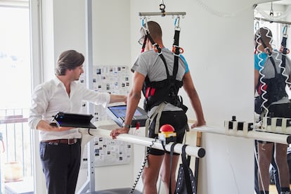
In a paper published in Nature, neuroscientist Grégoire Courtine of the Swiss Federal Institute of Technology in Lausanne and neurosurgeon Jocelyne Bloch of Lausanne University Hospital described how a system developed for rats was fine-tuned and refined into an implant that is placed in the lower part of a human spinal column where it emits electrical stimuli. Since the patients’ spinal cord injuries cut the lower body’s connection to the brain, a tablet was used to issue wireless signals that the lower body usually receives from the brain. The astounding result was that nine severely injured people were able to walk again with walkers and even crutches.
To work out why it was so effective, like the Swiss clockmakers of old, the neuroscientists took the whole system apart and put it back together again. Using various imaging techniques, they measured the metabolic activity of the medulla while the nine test subjects engaged in physical activity. Under normal conditions, the human metabolism accelerates during exercise. But after electrical stimulation of the spine, the neuronal activity of the test subjects actually dropped. Something unexpected was happening with the neurons in this part of the nervous system. To figure out why, Courtine and Bloch replicated all of their work – protocols, implantation, stimulation – in a group of crippled mice.
Working with mice was not easy. Besides the difficulty of adapting everything to the much smaller animals, the scientists counted almost 21,000 neurons in the rodent’s medulla. They then began the process of mapping the neurons and grouping them by genetic affiliation or function. Using optogenetics, a technique that uses flashes of light to activate and deactivate neurons, they were able to develop a complete 3D neuronal atlas of the medulla. They found that a special type of neuron – the VSX2 interneuron – was stimulated by EES. But this only happened with the crippled mice, not the healthy ones. Better yet, the activity persisted even when the EES was turned off. Eureka! They had just confirmed that the brain’s neuronal plasticity can also be observed in the spinal cord. Moreover, they had identified a group of nerve cells with the capacity to change function after an injury or trauma.