How scientists are using ultrasound to control neuron functions
Research is paving the way toward the ‘sonogenetic’ era, via the manipulation of mouse neurons using sound waves
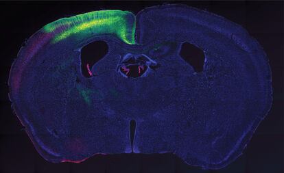

A group of researchers has managed to manipulate the neuron functions of mammals using ultrasound. To do so, they used a protein that is sensitive to high frequencies. After genetically introducing it into the brains of mice, they managed to move the muscles in their limbs. They repeated the experiment with human cells, and also managed to activate them, according to research published on the Nature Communications website last month. In the future, the researchers say, sonogetic control could be used across species. First, however, technical and ethical problems will have to be addressed.
Nearly a century ago, biologist Edmund Newton Harvey discovered that the resected heart of a frog would start beating when ultrasound was used. Since then, these sound waves – which can’t be heard by humans – have been used for ultrasound scans and little else. Among their limited clinical uses are the treatment of neuropathic pain and last-resort surgeries for cancer cases. Despite being a non-invasive procedure, ultrasound techniques had not, until now, managed to solve their biggest problem: their low precision at a tissue level.
The solution to this problem lay with a worm. In 2015, a group of scientists from the Salk Institute in the United States discovered how to manipulate the individual neurons of a Caenorhabditis elegans, a roundworm that is often used in research. They identified a protein that was sensitive to ultrasound, and saw that this protein stopped activating when it received the soundwaves. By doing so, they managed to change the movements of the worm. This team, led by Sreekanth Chalasani, is the same one that has achieved something similar but this time in mammals.
The task did not look simple. The nervous system of the C. elegans, as the roundworm is also known, only has 302 neurons and all of its synapses are known. Humans have more than 100 billion neurons and there is still much of the synaptic tree to be mapped out.
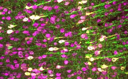
The first thing that they did was to look for the ultrasound-sensitive protein from the worm in mammals, but they couldn’t locate it. They also tried to register it in the cells of vertebrates, but also came up short. So they had to analyze some 300 different proteins by looking for one that was sensitive to ultrasound. And they found one: it’s called TRPA1 and belongs to a group TRPs, which work like channels of the cellular membrane, opening and closing paths to the inside of or from the cell.
Spaniard Marc Duque Ramírez is the first author of this research. “Ultrasound makes the membrane vibrate, but the effect goes beyond the mechanical,” he explains. And that effect is to do with the lipids that comprise both the outer layer of the cell and the cytoskeleton, the protein structure that support the cellular structure. “The channels open, and calcium ions enter – positive ions that generate the cellular response.”
The scientists modified the genes of mice to produce the human TRPA1 protein. Then they used an ultrasound transducer, which converts an electrical signal into high-frequency sound waves. They found that the neurons that specifically contained the protein were activated, but not others. The end result was a controlled movement of the limbs.
While the study focused on neurons, this could be used with cells in other organs. “I applied the ultrasound to cardiomyocytes [cells from the cardiac muscle] in mice and saw how they started to beat again,” explains Duque, who is now carrying out research at Harvard University. Another possible field that the Spanish researcher points to is that of the pancreas, “which could even induce an increase in the secretion of insulin.”
As well as the technical challenges, Marc Duque, one of the authors of this new research, believes that the ethical problems must first be solved. “There is no consensus nor protocols on the expression of foreign genes in human cells and even less so in the brain,” he explains. The issue of mind control immediately rears its head. But his then-boss at the Salk Institute when these proteins were discovered, Chalasani, plays down the possible reluctance and frames the debate. “On its own, ultrasound is not enough to activate the neurons,” he says. “We need to express a channel to do so. So we would have to introduce this channel in the neurons we want to control. Without the channel, ultrasound doesn’t work. What’s more, nor does it travel through the air, meaning that the effect would be limited to the animal that expresses the protein and that was in contact with a transconductor.”