Mapping a tiny piece of mouse brain opens up new path to understanding human intelligence
Information gathered in just one cubic millimeter of a rodent’s cerebral cortex will help unravel the complex neural networks behind cognition

Mapping a tiny piece of a mouse’s brain — barely a cubic millimeter of its cerebral cortex — has opened a never-before-explored path toward understanding the human mind. An international consortium has successfully mapped, with unprecedented detail, all the neuronal wiring and how brain cells are activated in this small section of a mammalian organ. The data collected, which represents the most detailed brain mapping to date, will help unravel the complex neural networks underlying cognition and behavior. This research is part of the MICrONS (Machine Intelligence from Cortical Networks) project, widely regarded as the most complex neuroscience experiment ever attempted. The initial findings were published on Wednesday in the journal Nature.
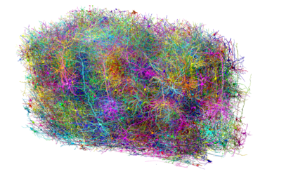
The tiny brain sample analyzed is no larger than a grain of sand, but it contains around 200,000 cells, 500 million synapses — the connections between neurons — and more than four kilometers of neural wiring.
“Within that tiny particle lies an entire architecture, like an exquisite forest. It contains all kinds of wiring rules we knew from various areas of neuroscience, and within the reconstruction itself, we can test old theories and hope to find new things no one has seen before,” said Clay Reid, a neuroscientist at the Allen Institute for Brain Sciences in Seattle and principal investigator of this project, in a statement.
Every idea, every memory, every action we perform in our daily lives originates from the activity of neurons in the brain — the intricate and enigmatic operations center that guards the human essence. Understanding how it works, how all the neural networks operate and relate to each other, and how each of their functions fits into the overall brain architecture is one of the greatest challenges facing the scientific community.
“Our intelligence and our mind are expressions of the physical structure of our brain. By understanding this structure, we can better define and shape hypotheses about how intelligence is implemented in our brain,” reflects Nuno da Costa, a scientist at the Allen Institute and co-author of this research, in an email response.
Brain wiring has already been successfully mapped in simpler animal models, such as the fruit fly larva (with just over 3,000 neurons and nearly 550,000 synapses) and the adult brain of the same animal (140,000 neurons and 50 million synapses). However, analyzing this tiny part of the mouse brain in such detail — which represents an even smaller fraction of the human brain — transcends, according to scientists, all known technological limits in the field of connectomics, the discipline that maps and describes neuronal connections.
Perhaps the only comparable effort, as two researchers from Harvard University note in an accompanying commentary, is the mapping of a cubic millimeter of the brain of an epilepsy patient last year: it contained 57,000 cells and 150 million synapses. “Together, these two projects [the mouse brain slice mapping and the epilepsy patient mapping] define the current technological frontier of large-scale mammalian connectomics,” the note states.
Da Costa says that, when they began their research, imaging a cubic millimeter of brain tissue with this level of detail “far exceeded” anything achieved to date. The experiment was, he says, “extremely ambitious.” “The scale and resolution of this dataset go far beyond neurons. It includes all blood vessels, non-neuronal cells such as glia, and even organelles within individual cells,” he adds.
He gives a visual example of what was achieved by profiling all those neural networks in the piece of mouse brain: “Imagine a kind of Google Maps for the brain: It will show not only the major highways, but also every street, every house, every room within every house, and even every door and window. Just as people use Google Maps to determine the best route from point A to point B, or even to check if a route exists, this kind of detailed brain map allows scientists to see if two neurons are connected and exactly where those connections occur.”
The region that makes us human
The volume studied is far from the size and complexity of a human brain, but the data from this mapping are more applicable than it might seem. The choice to analyze a specific region, such as the cerebral cortex, for example, is not trivial, explains Da Costa: “This brain region is possibly the most crucial structure that defines us as humans, largely due to its significant expansion in our brain. By studying how the cerebral cortex works in the mouse brain, we can gain better insights and hypotheses about how our own brain works.”
In the accompanying commentary, the two Harvard researchers elaborate on this: this brain region is considered “the seat of higher cognition,” a key territory for sensory perception, language processing, and decision-making. These are seemingly very different functions, but made possible by a kind of pattern found, with some modifications, in all cortical areas and in all mammals: “This makes studying the cortex in some ways similar to working out the principles of a combustion engine by looking at many cars,” states the commentary. “To understand the engine, it is useful not only to have a description of all its parts, but also to understand how the parts work together.”
The project was a tremendous team effort. First, scientists at Baylor College of Medicine in Texas used specialized microscopes to record brain activity in that cubic millimeter of a mouse’s visual cortex as the animal watched various movies and YouTube videos. Then, researchers at the Allen Institute took that same slice of brain and divided it into more than 24,000 layers, each more than a thousand times thinner than a hair, using electron microscopes to take high-resolution images of each slice. Finally, another group at Princeton University used artificial intelligence to reconstruct every cell and every connection in a virtual image, resulting in the largest wiring diagram and functional map of the brain to date.
More than 150 neuroscientists worked on the project. It required a titanic amount of work. “Cutting the more than 20,000 sections required for the data set took 12 straight days and nights, with our team working in shifts to ensure no consecutive sections were missed,” da Costa explains. A massive amount of information was collected — 1.6 petabytes of data, equivalent to watching an HD video continuously for 22 years. But their efforts have already begun to bear fruit.
“A giant step forward”
Early studies have revealed new cell types and innovative organizational and functional principles, explains the Allen Institute. Of note, for example, is the discovery of a new principle of inhibition within the brain. Thus, while it was thought that inhibitory cells (those that suppress neuronal activity) only dampened the action of other cells, researchers from the MICrONS project have discovered that the level of communication is actually much more sophisticated: inhibitory cells do not act randomly, but are highly selective about which excitatory cells they target and cooperate in networks (for example, some work together to suppress many excitatory cells, while others are more precise and target a single type).
And these discoveries are just the beginning. There are very high expectations about what insights this major research will yield. Not only will it advance our understanding of thought and consciousness, but it will also pave the way for progress in the study and treatment of numerous diseases.
“Brain diseases are ultimately the result of changes in brain structure, so understanding this structure is fundamental in the long term,” explains Da Costa. “In the medium and long term, detailed mapping of inhibitory cell connections, combined with genetic descriptions of these same cell types, could be crucial if any of these cells are found to be involved in a particular disease. This knowledge could, for example, support the development of drugs targeting specific subsets of the neuronal network or help explain how existing drugs work by identifying the cell types they affect.”
For Rafael Yuste, professor of Biological Sciences and director of the Center for Neurotechnology at Columbia University, who is the driving force behind the BRAIN Initiative, this research, in which he did not participate, is “a tour de force with a wealth of results, like laying one of many bricks in a huge building to understand the brain.”
“It’s worth remembering that more than a year ago, another batch of impressive articles was published, mapping the brain’s cell types, which are the neurons that generate all these connections. This is another of the most impressive results to emerge from the BRAIN project in the United States [the current research is also supported by the BRAIN Initiative]. Now, the great challenge is to bring together these two branches of new science; in other words, to understand what connections arise from what types of neurons,” the scientist said in statements to the Science Media Center (SMC) Spain website.
For his part, Juan Lerma, research professor at the CSIC-UMH Institute of Neurosciences, believes that this work “lays many of the foundations for several principles of functional organization that, although accepted, were not proven and represented gaps in our knowledge of the nervous system.” “These findings are a giant forward, long awaited, and are merely the tip of the iceberg of what is to come in understanding how the brain works,” he told SMC.
Sign up for our weekly newsletter to get more English-language news coverage from EL PAÍS USA Edition