Mini-brain cells created from human fetal brain tissue
Researchers have been able to study tumors in these 3D models measuring the size of a grain of rice, which had previously been obtained using isolated embryonic or stem cells
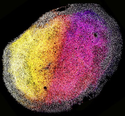

They are about the size of a grain of rice, but they are breaking new ground in research into the final frontier, the human brain. Dutch researchers have successfully created mini-brains capable of sustained growth from parts of the tissue of a human fetus. The study showed how these organoids included several cell types and expanded following a structure formed by proteins, also generated internally. This research offers a new way to study the functioning of the brain and, when appropriate, pathologies such as cancer in the organ that contains the human mind.
Traditionally, scientists have used different methods to model the biology of tissues, to study their function and that of the organs they form, as well as the associated pathologies. And they have done so by culturing cell lines, blood cells, liver cells, neurons and so on. Or using laboratory animals as models. For some years now, scientists have also been working with organoids, which are kinds of 3D mini organs with many of the morphological, developmental and even functional characteristics of the organ. This breakthrough is the result of the use of stem cells. Organoids can be formed directly from cells of a particular tissue, but researchers are also using pluripotent (adult) or embryonic stem cells to develop these into the organ to be studied.
Researchers at the Hubrecht Institute and the Princess Máxima Center for Pediatric Oncology (both in the Netherlands) have now developed a line of organoids not from individual stem cells, but from tissues of a human fetus donated for research after an abortion. In order to grow mini-organs, other approaches broke down the original tissue into individual cells. In this study, published in the scientific journal Cell, the team found that instead of working with portions of fetal brain tissue, they could self-organize by raising structures in three dimensions.
They can multiply in vitro, which means that from a small portion of fetal tissue we can generate multiple organoidsDelilah Hendriks, coauthor of the successful study
Dr. Delilah Hendriks, co-author of this investigation, explains in an e-mail what they have accomplished: “The fact that these organoids are derived from tissues means that we can study the developing human brain in vitro.” The study showed how proteins created by these cerebroids can be arranged to form extracellular matrices, the structures on which the rest of the brain cells multiplied until they became the size of a grain of rice. “Additionally, compared to existing models, they can multiply in vitro, which means that from a small portion of fetal tissue we can generate multiple organoids and these in turn can generate more, which is not only advantageous for reproducibility, but also makes it a powerful tool for genetic engineering, especially in the context of brain cancer modeling,” adds Hendriks.

That was the second part of their work. Using the CRISPR gene-editing technique, they introduced defects in TP53, a known cancer-causing gene, into several cells of the organoids. After three months, the TP53-defective cells had totally outnumbered the healthy cells. This enhanced ability to replicate is a typical feature of cancer cells. They then used the same gene-editing technique for the very opposite, to deactivate three genes linked to glioblastoma, the most aggressive of brain tumors. “We managed to deactivate all 3 genes at the same time. This means that we were able to introduce complete deletion mutations in TP53, PTEN and NF1 using CRISPR to engineer mutant organoids,” concludes Hendriks.
The organoids derived from the fetal tissues kept growing for more than six months, which allowed the scientists to continue to create many similar organoids from a single tissue sample. Meanwhile, mini-tumors with changes in the glioblastoma gene were also able to multiply, while maintaining the same combination of mutations. This could be used to perform repeat experiments with the same organoids, thereby enhancing the reliability and reproducibility of their results in future research.
“Brain organoids from fetal tissue are a valuable new tool for studying human brain development. We can now more readily study how the developing brain expands and observe the role of different cell types and their environment,” explains Benedetta Artegiani, leader of the Princess Máxima Center group, in a statement. “Our new tissue-derived brain model allows us to gain a better understanding of how the developing brain regulates the identity of cells. It could also help in understanding how errors in this process can result in neurodevelopmental diseases such as microcephaly, along with other diseases that can result from impaired development, including childhood brain cancer,” adds the researcher.
Professor Jacob Hanna, from the Pluripotent Cells and Ex Utero Embryogenesis Study Laboratory of the Weizmann Institute of Science (Israel) evaluates the results of this study: “It is important because it may offer clues about what occurs in brain development during actual brain formation, rather than relying on embryonic stem cell-derived tissues,” he told the SMC news department. Meanwhile, the researcher at the National Center for Biotechnology (CNB-CSIC), Lluís Montoliu, calls for caution: “Organoids are not the equivalent of the organs they model. This is neither in complexity nor in diversity of cell types. That is why we should remain cautious when interpreting the results that we can derive from the use of organoids in research.”
Sign up for our weekly newsletter to get more English-language news coverage from EL PAÍS USA Edition