Three new atlases reveal a new dimension of the human body
An international consortium analyzes the cell-by-cell architecture of the placenta, intestine and kidneys, moving closer to achieving the dream of creating the first map of the entire organism

Each person is an ensemble of 37 trillion cells — a number higher than the amount of galaxies in the entire universe — working in unison to keep us alive and healthy. Understanding how this enormous cellular orchestra coordinates to play the same symphony is one of biology’s greatest mysteries.
Three reference atlases were published this Wednesday, revealing this coordination in three organs that are essential for life: the placenta, which allows a fetus to grow in its mother’s womb without being destroyed; the kidneys, which cleanse the blood; and the intestines, which provide essential nutrients to the rest of the body. These three new atlases not only describe the human body cell by cell and specify them by type, but the maps also show how the cells are grouped and related to each other to form the detailed architecture of each tissue. It is a new dimension of the human body that may provide additional clues for understanding health and disease.
The new results are the product of five years of work by the Human Biomolecular Atlas consortium, which is composed of 400 scientists and generously funded by the U.S. National Institutes of Health, the largest public biomedical research organization in the world; the United Kingdom and Switzerland are also participating in the research project.
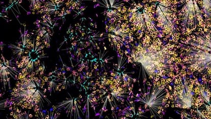
A year ago, the first results from the Atlas of Human Cells, a more international project with 3,000 researchers from 97 countries, were made public; that was the first major attempt to visualize the entire human cellular orchestra. That project produced the first image of the complete cellular orchestra and revealed how many instruments there were: some 500 different types of cells; it was previously thought that there were only 300. Now, the atlas provides information about where each musician sits, how they relate to their neighbors and how they are distributed by group. These results, derived from analysis of over two million cells from dozens of people, were published this Wednesday in the leading scientific journal Nature, and in six other complementary studies, which also include the first atlas of the skin.
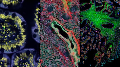
Geneticist Michael Snyder’s team used nine intestine samples taken from deceased people who donated their organs prior to their deaths. The researchers analyzed the three-dimensional cell-by-cell composition of eight parts of the intestine along its seven meters in length. A new technique called CODEX allowed them to sequence the proteins in each sample and obtain their spatial coordinates. The results reveal that different cell types, such as the epithelial cells that line the inner walls of the digestive tract and the immune cells that fight unwanted invaders, form “neighborhoods” with specific functions. In turn, these neighborhoods are grouped into “communities” with more distinct cell types, which collaborate with each other and determine the proper functioning of each part of the intestine.
These groupings vary according to a person’s health. For example, overweight or obese people’s cell communities have far more macrophages, immune cells that cause inflammation. The intestinal neighborhoods of people with hypertension also differ from those of healthy individuals.
Washington University physician Sanjay Jain completed the atlas of the human kidney using 45 samples from healthy people and 48 samples from those suffering from kidney disease. Researchers identified over 50 types of healthy cells, several of which were previously unknown to scientists; another 30 kinds of diseased cells, affected by acute and chronic ailments; and other cell subtypes that are in the process of recovering from external or internal damage. “We are looking at the interaction of neighborhoods of healthy cells and how they change with disease, which will likely help us design new therapies,” Jain explained at a press conference.
Michael Angelo’s team at Stanford University prepared the atlas of the tissue that connects a mother’s body with her unborn child’s, employing samples from 66 healthy women who decided to have an abortion between six and 20 weeks of pregnancy.
This third atlas enables the observation of a very difficult-to-access critical moment in human development with unprecedented clarity: the moment at which an embryo is implanted inside the mother’s uterus and her arteries begin redirecting to deliver oxygen and nutrients to the fetus, all thanks to a new organ, the placenta.
Humans experience enormous blood vessel reconduction compared to other mammals, even other primates; scientists do not know why that is the case. It could be an evolutionary adaptation to allow us to walk upright while maintaining sufficient blood flow for the fetus’s brain and other organs to develop properly in the last trimester of pregnancy. In turn, this phenomenon makes humans more prone to preeclampsia, one of the most common complications in pregnancy.
The placental atlas reveals amazing cellular relationships. Natural killers from the maternal immune system, cells that are designed to destroy all invaders paradoxically shift to favoring the fetus and ensuring that the immune system tolerates it, despite being an intruder from the genetic perspective. This new level of understanding makes it possible to explore and avoid reproductive problems, as well as similar phenomena, such as the rejection of transplanted organs and the growth of tumors.
Mapping the human body cell by cell will still take a long time. The U.S. project, which is now at the halfway point, seeks to produce detailed atlases of at least 30 of the nearly 78 organs that exist, according to the most widely accepted estimate. That data will then feed the global Human Cell Atlas project, explains Aviv Regev, who was one of this initiative’s founders in 2016 and is now the head of research at the U.S. drug company Genentech. “These new reference atlases are of great use to all researchers of health and disease because they give us a better understanding of the different cell types in these tissues,” she says. The researcher highlights another very recent achievement: the reference atlas of human lungs, which was created after analyzing 2.5 million cells from 486 donors of different ages, health statuses and geographic origins.
“Until now, the cell atlas was similar to using Google Earth in 2006 to look for your neighborhood and a blurry spot appeared,” explains Miguel de Oliveira Monteiro, the director of functional genomics at the Institute for Research in Biomedicine in Barcelona, Spain. The work published today uses new spatial genomics techniques that make it possible to give coordinates for each cell within the tissue and to see in real time which proteins, DNA and RNA they use. “Because of these new techniques, we’re seeing everything in detail for the first time… They show us the way forward to better understand diseases like cancer and processes such as aging, [and help us] comprehend the dysfunctional and the pathological,” Oliveira explains.
Creating the first human cell atlas represents an unprecedented scientific challenge. First, it was necessary to agree on whether the atlas should be done of a single person or many people, and then researchers needed to bring together the atlases of each organ, which are made using different techniques. In addition, a consensus had to be reached on how many types of cells exist in each one. The result will be a modern-day Frankenstein. Analyzing the data, doing the programming and computing necessary to reconcile it all, and creating the artificial intelligence to manage everything, all require a tremendous amount of work.
Holger Heyn, the director of single-cell genomics at the National Center for Genomic Analysis in Barcelona, Spain, heads the Human Cell Atlas’s technology unit and led another recently completed mapping: an atlas of the tonsils, which are the body’s first line of defense against pathogens that enter through the mouth. Researchers have identified 121 different cell types in this organ alone. “Between this year and next, we hope to have atlases of most of the organs. Then comes the biggest computational challenge: integrating them all into a single body; by definition, that will include the entire world, because we work with samples from most countries in the world. In two more years, we may have the first global model,” he says.
Computer expert Katy Börner, who is responsible for the U.S. project’s computational platform and a leading international expert on scientific maps, speculates on what the new global cell map will look like. “It will be an atlas that describes the male and female human body over its entire latitude and longitude, using the same frame of reference; that is a huge challenge given the enormous differences in size and function of the organs and the different techniques with which we are mapping them.”
Sign up for our weekly newsletter to get more English-language news coverage from EL PAÍS USA Edition