This is how the brain directs our movements
New research reveals that the organ’s control of voluntary actions is much more complex than we had imagined

Do me a favor: put your hands in front of your eyes and stop for a moment to look at them in detail from both sides. You will quickly appreciate that they show a special, authentic, and even beautiful design. They are one of the great achievements of biological evolution. The fingers, adjusted in number and size, have a great ability to carry out tasks and penetrate inaccessible, remote places. The thumb, by being placed in opposition to the other fingers, turns what would be a simple shovel into a powerful grip. The list of what we can do with our hands, such as moving things that we need or that are in our way, putting food in our mouths, getting dressed, using tools, writing, driving a vehicle — to name just a few examples — is endless.
But that’s not all, because the hands, in addition to being a powerful instrument to generate pleasure (massages, caresses, sexual stimulation), have a special ability to greet and express agreement and disagreement, as well as desire and feelings. That’s why we often say that hands “speak,” that they communicate. Babies, like our prelinguistic ancestors, can communicate with their arms and hands long before they do so with their larynx. The hands came into the world to revolutionize it. If we approach the world with our legs and feet, with our arms and hands we bring the world closer to us, we adjust it to our needs and purposes. Without them and without hands, the great technological and scientific development of our modern societies would not be conceivable. They’re a development that today more than ever controls the biological evolution of the brain and the fate of our species.
The way in which the brain controls the voluntary movements of the hand and other parts of the body began to be investigated in 1928, when the neurosurgeon Wilder Penfield, founder and first director of the prestigious Montreal Neurological Institute, tried to remove abnormal areas of the brain as a remedy to cure epilepsy. To this end, he developed a methodology based on electrical stimulation that allowed the anatomical identification of those important areas of the cerebral cortex, such as the areas related to language and movement. Injury to these areas could cause even greater disorders in patients than those that were intended to be eliminated.
In a typical brain operation, Penfield performed a cranial trephination under local anesthesia and exposed the brains of his patients. The patients remained conscious throughout the operation. In this way, he could move his electrodes from one point to another in the cerebral cortex, observing the influence of electrical stimulation on the movements and behavior of the patients. In hundreds of operations, his team collected data that enabled the localization of various sensory and motor functions in the pre- and postcentral gyri of the cerebral cortex.
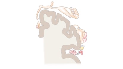
Following this method, they established a continuous cortical map, that is, a strip of nervous tissue whose neurons, organized in the same spatial order as the different parts of the body, control the voluntary movements of each part of the body. This map can also be represented by drawing a little man (the motor homunculus), whose limbs and body parts are of a size and proportions that, rather than corresponding to their real size, correspond to the proportion of cerebral cortex devoted to the movement of each one of them.
Thus, in the motor homunculus, the highly mobile parts of the body, such as the hand and its fingers, particularly the thumb, are large, while other parts with less mobility, such as the legs and feet, are smaller. In short, it’s a drawing that shows us the parts of the body with the greatest motor capacity as they relate to greater control of the cerebral cortex. Beyond this strip, other nearby regions of the frontal cortex have also been discovered that are involved in creating the spatiotemporal sequences of neuronal activity that allow complex voluntary movements to be carried out.
But that model of cortical simplicity (particular neurons controlling the movements of particular parts of the body), which is what we have been teaching in universities and research centers for many years, has been progressively questioned by the results of new research based on modern technologies. In the excellent and detailed work that has just been published in the scientific journal Nature, a large research group from several universities in the United States used high-precision functional magnetic resonance techniques to show that, instead of being continuous, the motor and classical homunculus is broken up by regions that have distinct connectivity, structure, and functions.
In other words, the motor cerebral cortex is divided into alternate regions for different functions. Up until now, three motor regions representing the foot, hand and mouth have been well recognized, but between them there are three other very different regions, called intereffectors, which are functionally interconnected and coupled to another annexed group of cortical regions (cingulo-opercular network) involved in mental control (preparation and implementation) of motor actions. These newly discovered regions are also seen in macaques and in young children, suggesting that this is an evolutionarily conserved primitive brain organization, originating early in infant brain development.
In short, as we should have guessed, the brain control of voluntary movements is anatomically and functionally much more complex than we had imagined until recently. It is not as precise or as continuous and linear as we thought; and there is still a lot to know before we can have a detailed vision of how the brain works, so that we can execute the voluntary actions that we require.
Sign up for our weekly newsletter to get more English-language news coverage from EL PAÍS USA Edition