AI model finds facial clues to estimate mortality risk in cancer patients
The system, trained on more than 40 million photographs, found a link between looking older than one’s actual age and a poorer prognosis

There is no proof that the face is a window to the soul, but a growing body of research suggests it may reveal important clues about a person’s health. Some studies have even indicated that looking younger than one’s actual age is associated with greater longevity. A new study published in The Lancet Digital Health follows this line of inquiry through a massive artificial intelligence analysis showing that facial photographs can contain signals linked both to age and to survival among cancer patients.
To reach their conclusions, the researchers trained the system on more than 40 million facial images and developed two tools from that analysis. The first estimates a person’s age. The second ranks cancer patients according to their risk of death. For now, however, the model is preliminary and still has a long way to go before demonstrating genuine clinical usefulness.
The age-estimation system, dubbed FAHR-FaceAge, was trained on 743,152 portraits for which the subjects’ true ages were known. It was then tested on a separate set of photographs it had never seen before. When estimating a person’s age, it had an average error of 5.1 years. When the researchers applied the method to 4,892 patients undergoing radiotherapy, the average discrepancy rose to nearly seven years.
The authors interpret this gap as a possible indicator of biological aging. If the AI estimates that a person is 70 years old when they are in fact 60, it suggests that their biological age may be 10 years greater than their chronological age, suggesting accelerated aging.
The second part of the study found an association between this age gap and survival among 38,183 cancer patients treated with radiotherapy. Patients who appeared at least five years older than their actual age had a 21% higher risk of death than the others.
In a second algorithm, FAHR-FaceSurvival, the researchers trained the system using 34,363 photographs of cancer patients together with their survival data. After adjusting for variables such as age, sex, type of cancer, treatment intent and radiotherapy technique, the quartile classified as being at the highest risk had more than three times the mortality rate of the lowest-risk quartile.
Raymond Mak, a radiation oncologist at Mass General Brigham and one of the study’s lead authors, suggested a potential use for the technology in assessing older or frail patients before recommending intensive treatments: “Our goal is to make health assessment from a photograph more quantitative, consistent and scalable, so it can support personalized decisions, not replace medical judgment.”
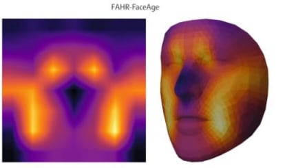
The authors found that the system’s age estimates varied little with makeup, lighting, facial expression or photo angle. They also observed that the age-prediction model focused primarily on the nasolabial folds, forehead and temples, whereas the survival model paid greater attention to the area under the eyes and the bridge of the nose.
What these observations do not provide, however, is a biological explanation for the gap between chronological age and facial age. “The fact that we are predicting survival is not enough to validate a measure of aging,” warns Leonor Cerdá Alberich, co-head of the Biomedical Imaging Research Group at La Fe Health Research Institute.
“We would need to verify whether that difference is related to indicators known in the literature on physiological aging, with covariates such as diseases or factors that contribute to appearance. The association with mortality is there, but we don’t know why. It remains unclear whether it responds to a specific biological signal, to disease, to habits, or to a combination of all of these,” she adds.
To answer that question, the facial-age estimate would need to be compared with biological-age biomarkers already used in research and clinical practice, such as epigenetic clocks based on chemical modifications to DNA, inflammatory markers, immune function, muscle strength and cardiorespiratory fitness.
Manel Esteller, a researcher at the Catalan Institution for Research and Advanced Studies (ICREA) and head of the Cancer Epigenetics Group at the Sant Pau Research Institute, also believes that “many follow-up studies confirming the findings” will be needed before the study’s prognostic claims can be considered reliable. Still, he notes that physicians have always used the face as an initial source of clinical information: “A yellower complexion may reflect a liver condition; very swollen eyelids, a kidney problem. With the arrival of artificial intelligence, that artisanal process has advanced by several orders of magnitude.”
The scientific literature already contains several studies that point in a similar direction. Esteller’s group published a study in 2022 showing that people with similar faces (virtual twins) also tended to have similar genomes, suggesting a link between facial features and the genetic predisposition to develop certain diseases. But detecting such patterns and turning them into a reliable clinical test is still a long way off.
Cerdá believes the main strength of the new study lies in its scale: “It is a first step, and provides promising evidence on the prognostic value of facial information. We can consider that a photo might be useful because it is associated with mortality or survival. But we still need to know how to interpret that signal and what it adds beyond what we already know.”
Before such a model could be applied in real-world clinical practice, the researcher continues, its predictions would need to be tested against the information already provided by clinical guidelines for cancer care, which are based on factors such as tumor stage, histological and genetic characteristics, the patient’s functional status, comorbidities, laboratory results and responses to previous treatments.
“In oncology, it is not enough to know that a variable is related to survival,” says Cerdá. “The question is what diagnostic gain it offers when added to what is already used. One would need to build a model with age, genetics, histology and all the clinical variables from the guidelines, add the image information and check whether it improves performance by 5%, 10% or not at all. The study does not address that comparison.”
The authors themselves acknowledge this limitation in the paper and call for prospective trials to test the tools alongside standard prognostic models. They also note that their cohorts are drawn primarily from high-income countries, that some racial groups are underrepresented, and that the risk of demographic bias remains.
Even so, the study marks a step forward in the use of the face as a measure of health and survival. Nothing similar could ever be demonstrated for the supposed qualities of the soul: attempts to link facial appearance or skull shape to moral virtues (or flaws) have long been exposed as pseudoscientific nonsense.
Sign up for our weekly newsletter to get more English-language news coverage from EL PAÍS USA Edition