Synchronization between the body’s circadian clocks can prevent aging
An experiment in mice reveals that a lack of coordination between the brain’s central timekeeper and the muscle’s molecular clock accelerates muscle tissue dysfunction. Reestablishing these communication networks helps restore proper functioning

Human life is governed by a circadian rhythm (around 24 hours) that is controlled by a tiny biological clock located in the brain. Based on the light stimuli entering through the retina, this molecular device synchronizes itself and tells the rest of the organism the time so that it can act accordingly. Night and day are not the same, not for the eyes, the liver, the skin or the pancreas. The peripheral clocks — located in organs and tissues — receive this instruction from the central chronometer and regulate themselves to set in motion one function or another, depending on the time of day. Like a kind of orchestra in tune, all these molecular instruments that manage the circadian rhythms communicate, interact and work, in turn, with the necessary autonomy to make the organism function. This is how the gears of life work.
If those clocks that mark the rhythm of existence did not exist, aging would accelerate. This has been seen in mice: in functional studies, when animals were created without these molecular chronometers, they aged prematurely and died much earlier, as if a human died at age 40. In practice, the mice had all their genes, the capacity to express them correctly and perform their usual functions, but without these circadian clocks, they did not know the best time to perform these functions and the whole vital infrastructure ended up collapsing sooner rather than later.
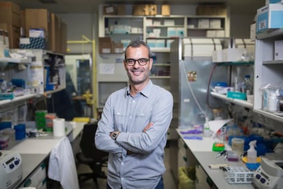
These tiny chronometers are key to survival, but their modus operandi remains, to a large extent, a mystery: the scientific community knows they play a key role in the vital process, but is still trying to unravel how exactly these communication networks are configured between one another. New research published in Science and Cell Stem Cell by Salvador Aznar-Benitah, head of the Aging and Metabolism Program at the Barcelona Institute for Research in Biomedicine (IRB), and Pura Muñoz-Cánoves, researcher at Pompeu Fabra University, who recently joined the multinational firm Altos Lab, has shed further insight on how these molecular clocks interact with one another. In experiments with arrhythmic mice, the study showed that a lack of coordination between the brain’s central chronometer and the one that regulates timing in the muscle accelerates the aging of muscle tissue. Restoring these communication networks, however, makes it possible to restore the function of this area and preserve its activity.
This is the first time the researchers have successfully tested in animal models a hypothesis they have been forging for more than a decade: the idea was that, in order to maintain circadian rhythms, each tissue probably has an autochthonous rhythm, which is independent from the rest of the organism. And that there is another process of interaction with the clocks of other organs to synchronize functions. “It makes a lot of sense that if our circadian rhythm is preparing us for food, the tongue, gut, pancreas and liver are all synchronized to know that they’re going to have to start metabolizing food. Imagine the problems that could arise if the liver gets ready at 2 a.m. and the stomach at 1 p.m.,” explains Aznar-Benitah.
In the study published in Science, the researchers designed an arrhythmic animal model — with deficiencies in the central clock, the muscle peripheral clock, or both — in order to dissect which circadian functions were performed by the tissue independently and which depended on communication with other clocks.
“The deregulation of our clock is one of the clear characteristics that happens to all of us as we age. What we saw during aging is that the clock machinery, the basic one, the one that tells the tissue that it is this time or that time, that does not change. So if we wanted to find possible therapeutic ways to keep the clock in a young state in the old organism, we had to understand what happens to the clock. And what it is telling us is that a large part of what happens to the clock is not that the machinery isn’t working well, but that the synchronization with other tissues, both peripheral and central, are what have changed. And we had to understand in which part of the functions the tissue does not need communication, and in which part of the functions it does need it and with whom,” explains the scientist.
The experiment showed that in some daily functions, muscle tissue does not need to synchronize. “If you have an animal that does not have the clock except in the muscle cells, that muscle is capable of maintaining between 10% and 15% of its functions temporarily,” explains Aznar-Benitah. “What is basic, remains. And we think that there is an evolutionary advantage in that, because if all the functions of all the tissues were linked to one communication, if a person has an infection in the liver, there would be domino effect: if the liver fails, everything else would fail. The fact that these functions have been separated from the need to communicate and synchronize with others means that, even if a person has a heart problem, the skin maintains its ability to have a barrier,” says Aznar-Benitah.
The research also revealed that there another 30% or 35% of muscle tissue functions that depend on the central clock. “Between the 15% independent functions and the 35% that depend on brain interaction, we have already mapped half of the tissue functions. There are another 50% of functions that we know are circadian, but we have not yet identified with what the muscle has to communicate for this function to occur when it has to occur,” admits the researcher.
Calorie restriction to strengthen communication
The study confirms that the coordination between the molecular clocks of the tissues is “crucial” to maintaining the general health of the organism. In fact, experiments to reestablish communications between these body clocks improved the condition of muscle tissue. One mechanism studied was to subject mice to temporary caloric restriction — they only ate during the active dark phase (night feeding) — which researchers discovered “could partially replace the central clock and improve the autonomy of the muscle clock.” Circadian restoration through caloric restriction mitigated muscle loss, impaired metabolic functions, and decreased muscle strength in old mice. “Eating like this strengthens the communication” between the brain clock and the muscle clock in mice, says Aznar-Benitah, although he clarifies that these findings cannot yet be extrapolated to humans — nor can the impact of practices such as calorie restriction.
The researcher points out that both the Science study and the one published in Cell Stem Cell — which studied the communication between the brain clock and the skin clock — are a step forward in understanding how these precise molecular devices work. But they still have no practical application. In fact, he predicts, it will be necessary to analyze tissue by tissue to see what the autonomous role of each clock is and how its coordination with other chronometers influences it.
“I don’t think that communication between brain and peripheral tissue will always slow down aging. There will be tissues that are functionally much more dependent on that communication, while others will be much more dependent on other peripheral tissues. But we have to test that one by one. What we do know is that in tissues and organs that we have been studying for a long time, such as liver, muscle, skin, there is a clear benefit of reestablishing communication between the peripheral tissue and the central clock,” he explains.
In the skin, for example, time is key: the internal clock of this tissue knows that the best time to promote cell division of stem cells and regenerate the skin is when it is not in contact with ultraviolet light, which is mutagenic. If cell division occurs when the skin cells are exposed to UV light, they can be affected by mutuations and errors.
“What’s more, because these cells are dividing, the mutations [they acquire from UV exposure] would be spreading to daughter cells, which would inherit that mutation. What the circadian rhythm does is separate these processes: it tells the skin cells not to divide while there is a peak of ultraviolet light,” says Aznar-Benitah. His study published in Cell Stem Cell, which analyzed this separation between DNA division and ultraviolet light exposure, revealed that if these communication networks between the central clock and the molecular timer in the epidermis are broken, cell division occurs at the same time as ultraviolet exposure.
A “federation” of clocks, not a “dictatorship”
Juan Antonio Madrid, professor of physiology and director of the Chronobiology and Sleep Laboratory at the University of Murcia in Spain, calls the research “beautiful and elegant because it describes many interactions and answers many questions” through “very interesting genetic engineering work.” “It is true that it is in mice, but it is interesting because it reveals to us how the body’s circadian system is not a hierarchical system, like a dictatorship, where the brain’s clock rules. It is more like a clock federation where everyone contributes.”
Sign up for our weekly newsletter to get more English-language news coverage from EL PAÍS USA Edition