An international study suggests that radiation from a childhood CT scan increases the risk of developing cancer
Experts explain that the danger is low but call for doctors to avoid abusing the technique and to adjust the doses insofar as possible
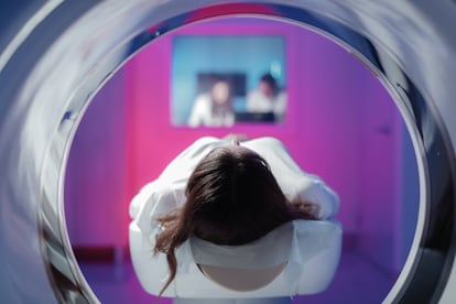

In medical contexts, radiation can be a double-edged sword. It is an extremely useful therapeutic and diagnostic tool for treating and curing many diseases, but it can also be dangerous to the body if the exposure or doses are too high: radiation causes mutations and changes in the DNA of cells that can cause cancer. Doctors who use this technique pursue a balance between the risks and benefits. Mindful of the risks, physicians are used to fine-tuning the guidelines and adjusting the doses to minimize the dangers and maximize the benefits. However, there is no such thing as zero risk when it comes to radiation. A new international study led by ISGlobal scientists in Barcelona, Spain, contributes additional evidence of the impact of radiation exposure, even at low doses: the research suggests that radiation from a childhood computed tomography (CT) scan increases the risk of developing a hematologic tumor. Published today in the journal Nature Medicine, the study estimates that, for every 10,000 children examined using this technique, one or two may suffer blood cancer in the subsequent 12 years as a result of this exposure. The experts emphasize that the individual risk is low, but their conclusions call for increased awareness among physicians not to abuse the technique and control exposure levels as much as possible.
Everyone is exposed to small amounts of radiation in their daily lives, from the ground, rocks, water, building materials… No one is spared from so-called natural background radiation. But in a medical context, the doses are usually higher than the levels of radiation in the natural environment. For example, a chest X-ray is the equivalent of one day’s exposure to natural background radiation; a cranial CT scan is similar to eight months of background radiation, and an abdominal CT scan is the same as about 20 months.
None of this radiation is harmless. The scientific community has long known that exposure to high doses of ionizing radiation is a risk factor for developing hematological tumors in both children and adults. For that reason, medical professionals closely monitor the guidelines and are careful not to overdo it; moderate doses are considered to be over 100 milligrays (the unit of measurement of the radiation dose absorbed by the body), while high doses exceed 1 gray. In fact, radiologists strictly follow the ALARA (as low as reasonably achievable) principle.
However, the actual risks to children and adolescents of exposure to radiation doses used in CT scans, which are typically below 100 milligrays, have long been “uncertain” and a subject of debate among scientists, the ISGlobal researchers note to explain the purpose of their research. “CT scans are procedures that have been used since the late 1970s and are a fantastic system for monitoring pathologies. With the development of this technique, there are increasingly more applications…15 years ago, we saw that its use had increased, and we decided to do a follow-up because [radiation] doses are higher than those of a normal X-ray. This study is not [intended] to cause alarm, but rather to assess the risks and generate data to ensure that patients are protected,” says Elisabeth Cardis, the head of the Radiation Group at ISGlobal and the study’s coordinator.
ISGlobal researchers analyzed data from nearly one million people from nine European countries who underwent CT scans before they were 22 years old. With the available information, the scientists attempted to reconstruct their radiological histories to estimate the radiation dose absorbed by their bone marrow. After cross-checking the data with mortality and cancer registries, researchers found an association between total bone marrow radiation doses from CT scans and the risk of developing blood cancer. The results showed that a current CT scan increases the risk of developing malignant tumors by 16%.
Cardis contextualizes these figures: “The risk is increased, but it is a low risk at the individual level.” She adds that the data falls within the expected range. “We did this study because, until recently, there was a debate about the effects of low doses. There were many radiation protection people who thought that nothing happened with low doses, and we questioned whether there was [no risk] or whether the risk was low,” she explains. The risk is small, but it cannot be underestimated.
The ISGlobal scientist says that, in recent years, pediatric protocols have been adjusted to reduce the doses used in pediatric patients and they have refined the guidelines for this type of medical test. But “there is still more that can be done,” Cardis says. “The [scan] can be optimized further to reduce doses and maintain good image quality. We advise that physicians and radiologists be more mindful: they should ask for a CT scan when necessary, but they should also look to see if there are alternatives.” Cardis gives the example of pathologies that require several CT scans in a short period of time to monitor a disease; in such cases, high-resolution images might not be needed to monitor the ailment and the radiation dose can be reduced.
‘There is no need for alarm’
Nevertheless, the researcher advises caution and rejects any alarm that her study’s conclusions might cause: “CT is an indispensable tool that is used because there are more serious health risks. CT saves lives. It is necessary [for doctors] to calculate the risk and the benefit because the benefit can be very significant, and the risk is decreasing because the doses have been significantly reduced. There is no need to worry too much.”
A few years ago, Ignacio Barber Martínez de la Torre, the director of Pediatric Radiology at the Sant Joan de Déu Hospital in Barcelona, Spain, participated in recruiting participants for the study and is familiar with the research, although he did not participate in this latest study. He observes that the findings add more scientific evidence to risks that are already known: “We know the risk and we know what happens with higher non-medical doses, due to nuclear accidents. That is why radiological protection measures have always been used… In children, there are two clear risks: they are more radiosensitive and there’s more time for them to develop diseases [after exposure].” However, the radiologist points out that current scan machines “irradiate much less” and says that the average radiation dose in this study is probably no longer the same as the one used now: “This cohort was chosen 10 or 15 years ago, and the technology has evolved a lot [since then].”
Josep Munuera, the director of diagnostic imaging at the Sant Pau Hospital in Barcelona (who was not involved in this research), also points out that the study “is very good methodologically and the high population size reinforces the quality” of the analysis. He notes that the study’s conclusions are in line with radiologists’ standard practice: “All scientific organizations advocate the ALARA principle: using the minimum dose because we know that X-rays have [both] a positive and a negative side, such as producing changes that can cause tumors. The value of this study is that it shows that this does not happen exclusively in cases where there’s excess radiation, but that it can [also] happen in patients who undergo a low number of tests.”
Munuera puts the issue into perspective: “We should not sound the alarm because we know this, but we must reinforce the idea of performing the most appropriate tests because doing imaging tests is not harmless.” The physician, who is also the scientific director of the Spanish Society of Medical Radiology, reminds us that the CT scan is a “basic and essential” tool, emphasizing that the new generation of devices already greatly reduce radiation. “The new generations of CT scan machines irradiate much less; [it is] almost equivalent to the radiation with an X-ray. The problem is repeated tests.” In any case, the radiologist adds that “we are governed by the ALARA principle, as well as the risk-benefit concept… If there is a diagnostic benefit, there’s a positive balance. And, lastly, the minimum dose must be used, but it must be diagnostic, because there is a physical limit at which the image is not diagnostic, and we need it to be sufficient to be able to evaluate.”
Speaking to the Science Media Centre, Sarah McDonald, the deputy director of research at Blood Cancer U.K., which researches the disease in the United Kingdom, also noted that this study is “large and well conducted” but “does not prove a direct causal relationship between a CT scan and the risk of blood cancer.” The researchers found an association but cannot prove causality: “Risk factors are not the same as causes, and there are several interrelated risk factors for blood cancer, and factors such as age, sex and ethnicity also play an important role,” McDonald said.
Barber Martínez de la Torre, who is also president of the Spanish Society of Pediatric Radiology, highlights the context in which these techniques are used, along with their importance. “Many of these tests are done in sick children, who have a life prognosis already shaped by the disease. Not using this technique can be a greater risk than using it. This publication reminds us that what we do, we do well, but we have to control it and improve… We have to be aware of these limitations and justify its use and limit the doses.”
Sign up for our weekly newsletter to get more English-language news coverage from EL PAÍS USA Edition